In December 2018, DIFFER received a desktop Thermo Scientific Phenom Pharos Scanning Electron Microscope (SEM). This small-sized SEM was one of the first of its kind in the Netherlands and it is now in regular operation at the institute. Due to its ease of use, the microscope speeds up DIFFER’s energy materials research significantly.
"Our researchers have always had the opportunity to use the Scanning Electron Microscope at the Nanolab@TU/e", says Stefan Welzel, group leader of of DIFFER’s Solar Fuels Facilities & Instrumentation group. "However, that specific piece of equipment is a specialized, room-sized microscope, which can only be operated by experts. Our new in-house desktop SEM is stripped of all the features we do not need, which makes it easy to operate. So now, even our students and interns can use it to quickly assess the morphology or composition of for example photocatalytically or electrochemically active materials."
Coffee machine to see nanometer-scale details
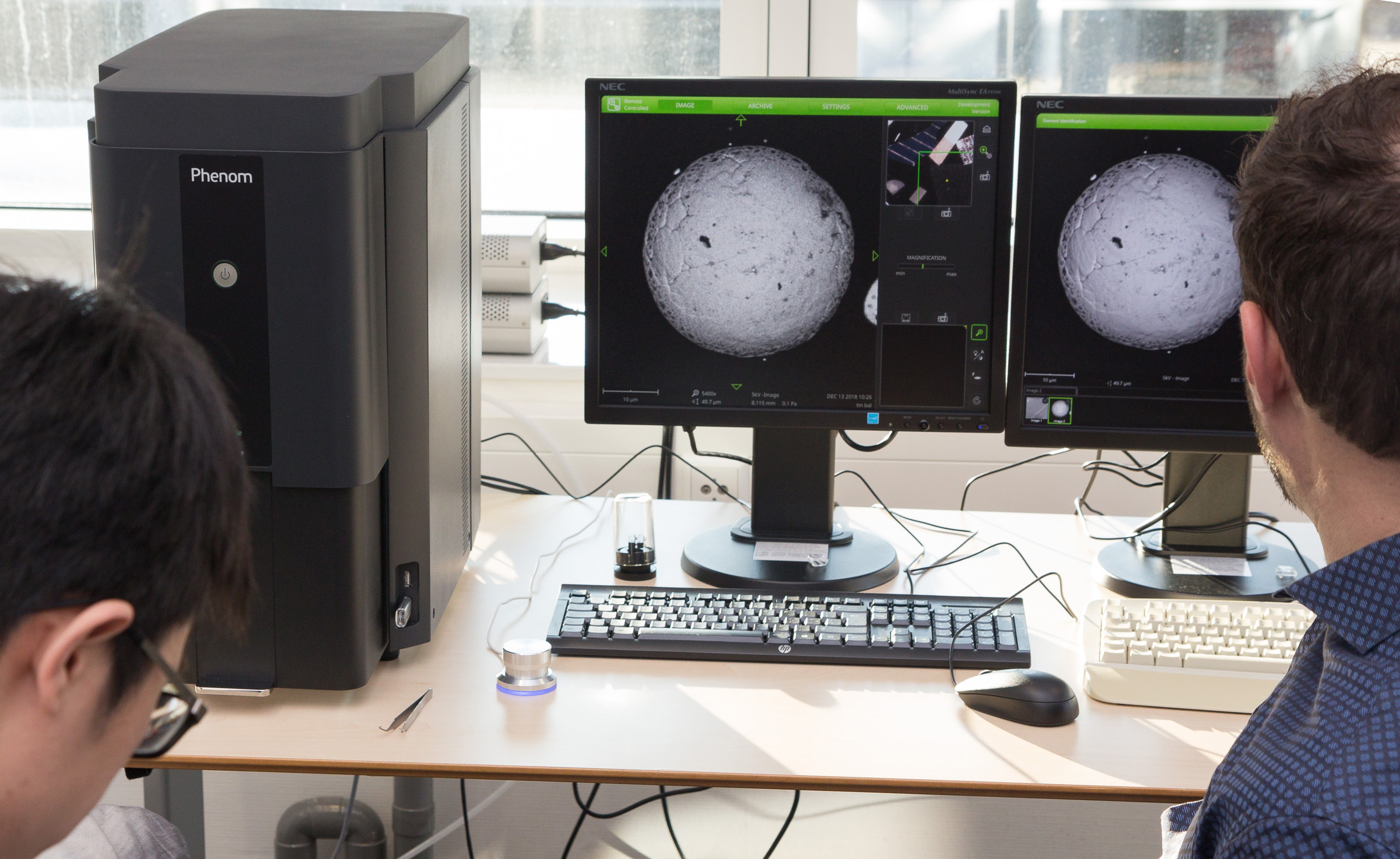
Though DIFFER’s desktop SEM is only as big as a coffee machine, it still enables the same resolution performance down to three nanometers that typical cupboard-size SEMs provide. "Another advantage of this desktop machine is that it doesn’t impose huge restrictions on the infrastructure, like the need to build vibrational-free floors. So when the SEM was commissioned to us in December, we could start using it almost right away."
In a Scanning Electron Microscope, a beam of electrons is directed toward a sample. By detecting both the backscattered electrons and the electrons that are knocked out of the atoms the sample is composed of, a 3D image of the surface of the sample can be made. In addition to that, DIFFER’s SEM is also equipped with an Energy Dispersive Spectroscopy function, which provides information about the chemical composition of the sample.
State-of-the-art characterization capabilities
Together with the Atomic Force Microscope and the Transmission Electron Microscope DIFFER had acquired earlier, the desktop Scanning Electron Microscope enables fast, state-of-the-art characterization of the morphology and composition of new materials for energy conversion, which are studied by research groups from the Solar Fuels and Fusion Energy themes at DIFFER.
Go to the News page.